
ファイル:Human brain left midsagitttal view closeup description 2-emphasizing-corpus-callosum.png
Human_brain_left_midsagitttal_view_closeup_description_2-emphasizing-corpus-callosum.png (702 × 491 ピクセル、ファイルサイズ: 1.32メガバイト、MIME タイプ: image/png)
ウィキメディア・コモンズのファイルページにある説明を、以下に表示します。
|
概要
| 解説 |
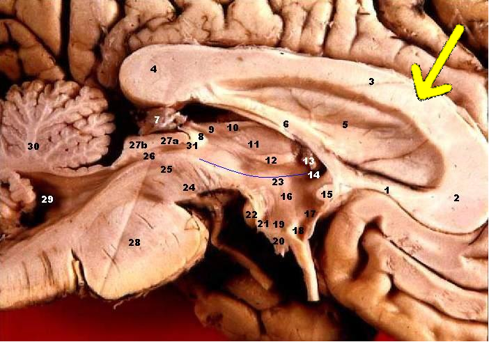
Human brain left - midsagitttal view - closeup. Emphasizing corpus callosum
On a half brain specimen, the Thalamus can be identified. The Thalamus (anteroom) is connected caudally with the midbrain and rostrally with the cerebral hemispheres. Note that the walls of the 3rd ventricle are completely formed by the Thalamus. The Hypothalamic Sulcus separates the Dorsal Thalamus superiorly from the Hypothalamus inferiorly. The subthalamus is located lateral to the hypothalamus and is not visible here. The anterior wall of the 3rd ventricle is a thin sheet of tissue called the Lamina Terminalis. In the Dorsal Thalamus note the Interthalamic Adhesion (Massa Intermedia), and the three parts of the Epithalamus: Stria Medullaris Thalami, Habenula, and the Pineal Gland. The floor of the Hypothalamus is made up of the Infundibulum, which connects with the pituitary gland, and posteriorly, the Tuber Cinereum and Mammillary Body.
|
| 日付 | |
| 原典 | http://www.healcentral.org/healapp/showMetadata?metadataId=40566 (Internet Archive of file description page) |
| 作者 |
John A Beal, PhD Dep't. of Cellular Biology & Anatomy, Louisiana State University Health Sciences Center Shreveport |
| 許可 (ファイルの再利用) |
CC-BY |
| その他のバージョン |
 |
ライセンス
- あなたは以下の条件に従う場合に限り、自由に
- 共有 – 本作品を複製、頒布、展示、実演できます。
- 再構成 – 二次的著作物を作成できます。
- あなたの従うべき条件は以下の通りです。
- 表示 – あなたは適切なクレジットを表示し、ライセンスへのリンクを提供し、変更があったらその旨を示さなければなりません。これらは合理的であればどのような方法で行っても構いませんが、許諾者があなたやあなたの利用行為を支持していると示唆するような方法は除きます。
当初、http://www.healcentral.org/healapp/showMetadata?metadataId=40566に投稿されたこのファイルは、2013年9月25日に画像査読者のEleassarによって査読され、その時点で、記載されたライセンスの下で利用可能であることが確認されました。
|
ファイルの履歴
過去の版のファイルを表示するには、その版の日時をクリックしてください。
| 日付と時刻 | サムネイル | 寸法 | 利用者 | コメント | |
|---|---|---|---|---|---|
| 現在の版 | 2009年10月1日 (木) 20:30 | | 702 × 491 (1.32メガバイト) | Was a bee | {{Information| |Description='''Human brain left - midsagitttal view - closeup'''. Emphasizing corpus callosum # Corpus callosum, Rostrum # Corpus callosum, Genu # Corpus callosum, Corpus # Corpus callosum, Splenium # Septum pellucidum # Fornix, Corpus # |
ファイルの使用状況
以下のページがこのファイルを使用しています: